Molecular nanocrystals for in vitro imaging or two-photon photodynamic therapy
The use of nanoparticles for biological imaging applications is booming, particularly for cancer diagnosis.
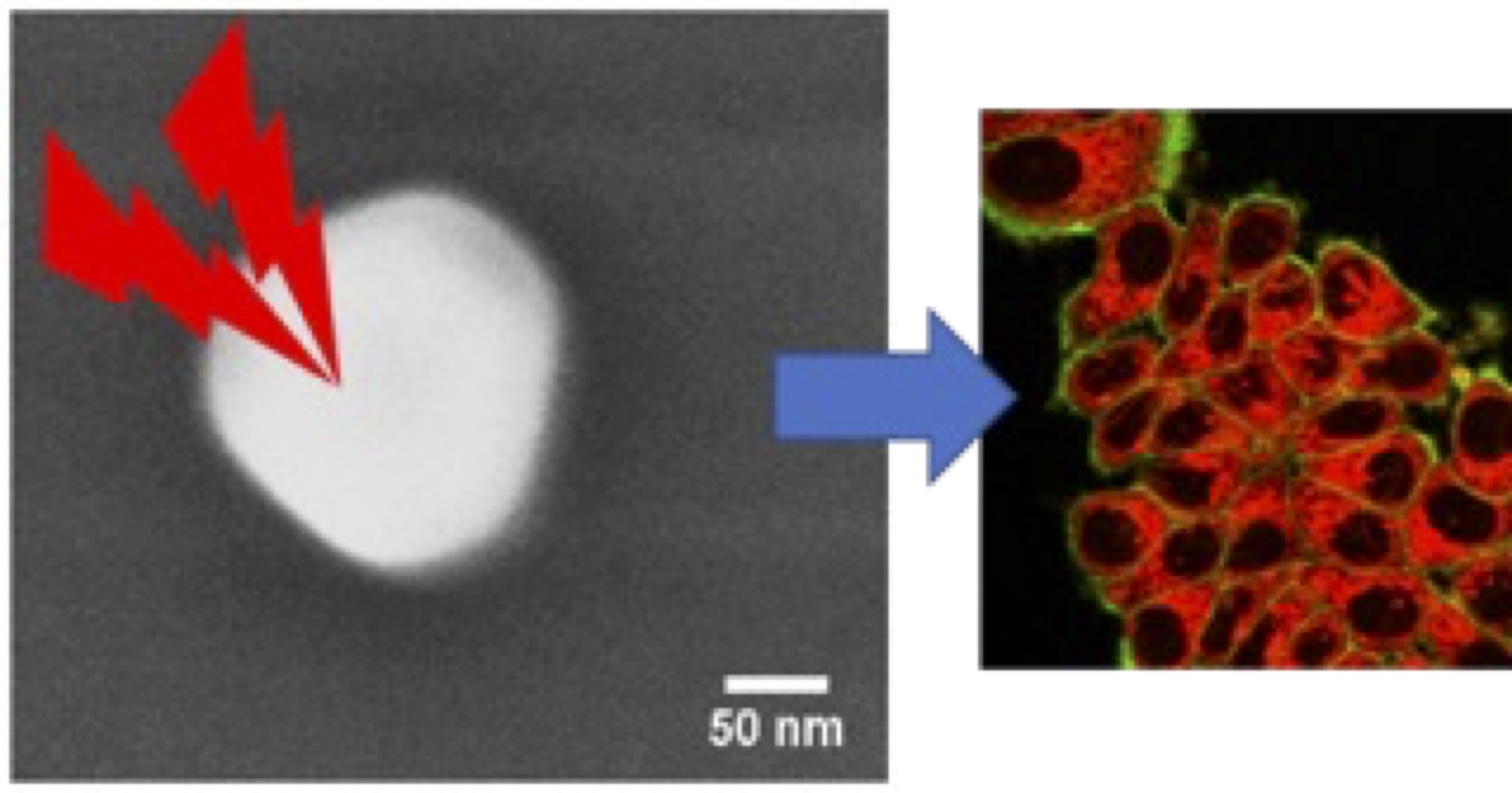
The use of nanoparticles (NPs) for biological imaging applications is booming, particularly for cancer diagnosis. In addition, these NPs can sometimes be used in photodynamic therapy if singlet oxygen is produced under irradiation. In particular, two-photon excitation of organic dyes enables in-depth imaging thanks to excitation in the near infrared, where tissue absorbs little, with ideally emission in the red or near infrared at the start of the first biological transparency window. However, as the probability of two-photon absorption is very low, the fluorophores in the NPs need to be concentrated as much as possible. The use of nanocrystals of organic fluorophores emitting in the crystalline state in the infrared and with two-photon absorption properties is therefore very promising. For a number of years, we have been studying a one-step process for synthesizing molecular nanocrystals coated with a silica shell using the spray-drying method. However, this method results in excessively high polydispersity, and the stresses exerted by the silica layer during its formation lead to the presence of numerous defects in the crystals, resulting in altered fluorescence properties. We have recently developed a sonocrystallisation set-up for preparing colloids of organic nanocrystals (50-100 nm). [1]
In this project combining crystallogenesis, nanochemistry, physical chemistry of colloids, fluorescence studies and applications in biology, we aim to optimize sonocrystallisation processes, develop the coating of organic nanocrystals with a silicate shell and their functionalization, and use these new types of fluorescent tracers for imaging or two-photon photodynamic therapy in vitro on cell cultures (in collaboration). In particular, we will develop nanocrystals of porphyrin derivatives known to be very active in two-photon PDT.
Particular emphasis will be placed on characterizing crystallinity (X-ray and electron diffraction) and photophysical properties.
Bibliography
[1]: Sonocrystallization of CMONS Needles and Nanocubes: Mechanistic Studies and Advanced Crystallinity Characterization by Combining X-ray and Electron Diffractions with DNP-Enhanced NMR
X. Cattoën, A. Kumar, F. Dubois, C. Vaillant, M. Matta-Seclén, O. Leynaud, S. Kodjikian, S. Hediger, G. De Paëpe, and A. Ibanez Crystal Growth & Design 2022 22, 2181-2191
Expected skills
The student should follow the Graduate School programm in Soft Nanosciences with the master: M1 Soft Matter and Biophysics, or M1 Nanochemistry, or Phelma Biomed Engineering.
Required: background in physical-chemistry. Strong interest in the development and characterizations of nanomaterials with spectroscopic properties
Published on April 12, 2024
Updated on April 12, 2024