Ultrasound localization and manipulation of air bubbles
Research project of Fidaa Qorom, student of the Soft Nanoscience program of GS@UGA, at the Liphy, Grenoble.
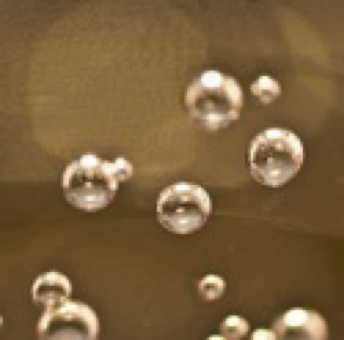
Biomedical ultrasound imaging is one the most widespread diagnostic modality. Many applications, such as obstetric imaging, are based on endogenous tissue contrast. However, ultrasound imaging may also benefit from contrast agents, which aim at enhancing contrast and/or specificity. Ultrasound contrast agents consist of encapsulated air bubble, with possibly very large scattering cross-sections with respect to their size. Not only can ultrasound contrast agents be used to enhanced ultrasound contrast [1], but they can also be functionalized to carry and deliver drugs, and enable super-resolution imaging [2]. Although ultrasound contrast agents have been used for decades [1], simultaneous imaging and manipulating contrast agents remains a key challenge for in vivo biomedical applications, where the contrast agents are carried by blood flow.
In this project, the main objective is to develop a proof-of-concept experiment in which air bubbles in a water tank are simultaneously localized and manipulated by use of an ultrasound array driven by a state-of-the-art ultrasound electronics.
Bibliography:
[1] Frinking, P., Segers, T., Luan, Y., & Tranquart, F. (2020). Three decades of ultrasound contrast agents: a review of the past, present and future improvements. Ultrasound in medicine & biology, 46(4), 892-908.
[2] Errico, C., Pierre, J., Pezet, S., Desailly, Y., Lenkei, Z., Couture, O., & Tanter, M. (2015). Ultrafast ultrasound localization microscopy for deep super-resolution vascular imaging. Nature, 527(7579), 499-502.
Published on April 9, 2024
Updated on September 4, 2024