Development on interferometric biosensors on optical fibers.
 Scientific context and objective
The objective of this internship is to contribute to the development of a diagnostic tool intended to eventually replace biopsies, long and invasive procedures. To achieve this goal, we propose to develop interferometric biosensors on an optical fiber assembly capable of performing real-time, remote, in situ, and multiplexed molecular analysis. Such a tool could have a dramatic impact in health technology, allowing faster (real-time) and less invasive analyses directly at the level of the diseased tissue, thus shortening the diagnosis time.
Biosensor principle
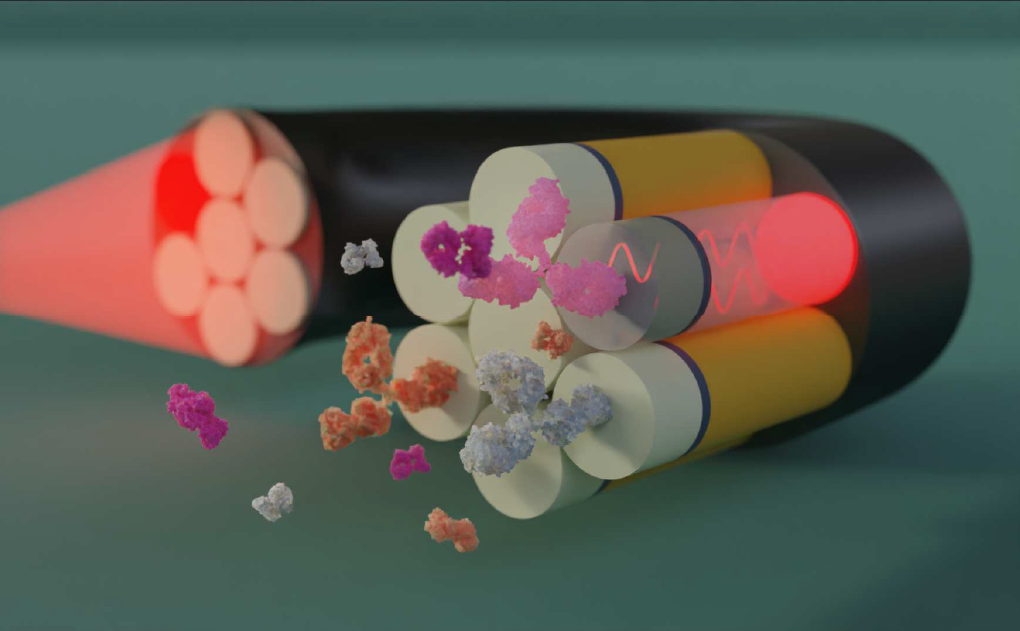
Based on our experience with fiber-based plasmonic biosensors, we have chosen to develop a sensing strategy based on interferometry. This label-free approach is particularly interesting for in vivo diagnosis. The figure provided below represents a schematic view of the tool under development. The detection principle is based on the interference between a light wave reflected by an internal reference layer and a second wave whose optical path is influenced by the interaction between targets and probes immobilized at the end of the fiber. This results in a real-time quantification of the interaction. Using a multifiber assembly, each fiber will act as an individual sensor, and the measurement of the intensity of the light retro-reflected by the functionalized side will allow monitoring of biological interactions occurring on this surface.
Trainee's work
Thanks to the modeling of the interference phenomenon occurring at the end of the fiber, we have determined geometrical parameters (thicknesses) and optical parameters (indexes) to maximize the sensitivity of the fibers to optical index variations. Surface treatments have been applied on different samples (single fiber, multicore fibers, multifiber assemblies...) selected to produce interferences. These sample have been characterized in terms of sensitivities to optical index variations and resolution.
In order to detect biological interactions, it is now necessary to functionalize the samples. The M1 trainee will start by testing different functionalization strategies on quartz slides using optical4 and chemical5 methods (photo-functionalization). For this, he/she will participate in the elaboration of biochemical grafting protocols and in the development of a new optical set-up dedicated to functionalization.
Thereafter, the established protocols and set-up will be adapted and implemented for optical fibers’ multi-functionalization (M2 internship). The detection limits for biological targets will be evaluated first using model probes and targets, secondly using targets of biological interest. The detection will be gradually done in more and more complex environments (serum, blood).
Bibliography
[1] Vindas, K., et al. Enhanced sensitivity of plasmonic optical fiber sensors by analyzing the distribution of the optical modes intensity. (2020) Optics Express Vol. 28, Issue 20, pp. 28740-28749. doi.org/10.1364/OE.399856
[2] Desmet, C., et al. Multiplexed Remote SPR Detection of Biological Interactions through Optical Fiber Bundles. (2020) Sensors, MDPI, Vol 20 (2), p.511. doi.org/10.3390/s20020511
[3] Vindas, K., et al. Highly-Parallel Remote SPR Detection of DNA Hybridization by Micropillar Optical Arrays. (2019) Analytical and Bioanalytical Chemistry, Vol 411, p2249-2259 doi.org/10.1007/s00216-019-01689-2
[4] Alvarado Meza, R., et al. Optically Assisted Surface Functionalization for Protein Arraying in Aqueous Media. (2017) Langmuir 33, 10511-10516
[5] Dendane N., et al. Efficient Surface Patterning of Oligonucleotides Inside a Glass Capillary through Oxime Bond Formation. (2007) Bioconjugate Chem. 18, 671 doi.org/10.1021/bc060254v
Opened to:
Student of the Thematic program Soft Nano in M1 Soft Matter and Biophysics, or Phelma Biomedical Engineering.Expected skills: Physics, optics, image treatment, surface chemistry/functionalization. Interest in biology and/or skills in molecular biology and/or biochemistry will be appreciated.
Updated on September 19, 2023