Role of peptide-insulin nanoparticles in insulin aggregation on hydrophobic surfaces
Research project of Ekaterina Imaikina, student of the Soft Nanoscience program of GS@UGA, at the LMPG Grenoble
Master research project in nanobiotechnology
Human Insulin (HI) is a protein produced in the pancreas, composed of two subunits totaling 51 amino acids. It is secreted in the bloodstream by Langerhans β cells and binds to cells having a specific insulin receptor (skeletal muscle cells, hepatocytes, adipocytes…). Diabetes is a frequent metabolic disease due to the destruction of Langerhans β cells (Type I) or decreased sensitivity of insulin receptors (Type II). All type I diabetic patients and about 10% of Type II diabetic patients require lifelong insulin supplementation. Insulin aggregation into amyloid fibers is a restricting factor to the development of embedded insulin delivery devices, since insulin tends to aggregate when exposed to hydrophobic surfaces such as catheters, forming amyloid fibers. Research for an excipient capable of limiting the fibrillation of insulin has been ongoing for years but no suitable candidate molecule has yet been found.
Insulin fibrillation kinetic is conveniently monitored using fluorescence-based assays. For instance, Thioflavine T (ThT) is a fluorescent conformational marker that binds only to amyloid fibers and red-shifts excitation and emission wavelengths. Insulin aggregation kinetic is divided in 3 phases. First a lag phase during which no ThT fluorescence is detected, then a growth phase where the ThT fluorescence increases linearly in time, finally a plateau where the fluorescence remains constant. Seed experiments unambiguously demonstrated that the formation of ThT-positive insulin aggregates takes place at the plastic surface, and that insulin fibers are later released in solution.
[Ratha et al., 2016] reported that small amphipathic polypeptides such as KR7 (KPWWPRR-NH2) inhibit the fibrillation process, up to 80%. However, the peptide concentration to achieve inhibition is comparable to insulin, precluding its use in therapy. [Chouchane, 2017] reported that repetitive peptides such as LK5 (LKLKL), LK7 (LKLKLKL), LK9 (LKLKLKLKL) and LK11 (LKLKLKLKLKL) strongly influence insulin aggregation at smaller concentrations (less than 1:100 peptide-insulin ratio). More precisely, these peptides activate fibrillation at low concentrations (0.1 µM) and inhibit it at high concentrations (5 µM). Practically, these two effects are dissociated in two-step experiments : the plastic surface is pre-coated with LK11 during 5 min, then washed with buffer to remove non-adsorbed peptides, then incubated with insulin. Aggregation starts after a short lag time (about 30 min). Adding 5 µM LK11 immediately slows down the aggregation rate.
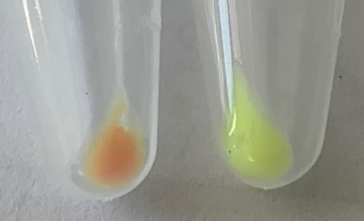
Recently, [Qu, 2022] showed that ThT-negative aggregates are released from the plastic surface into solution during the lag time. These aggregates were recovered by centrifugation. Using labelled proteins (FITC-Insulin) and labeled peptide (TAMRA-LK11) he was able to demonstrate that this protein material consists of co-agregated insulin and peptide (hybrid insulin-peptide aggregates) and quantify them. Furthermore, the spectral overlap of the two fluorescent dyes allows recording Förster resonance energy transfer (FRET) signals confirming that the fluorophores are separated by less than 10 nm. The existence of protein aggregates were confirmed by Dynamic Light Scattering (DLS). The initial starting insulin solution exhibits a single peak, at about 6-7 nm, whereas the liquid fraction recovered during the lag time exhibits a second peak at about 400-500 nm in addition to the 5 nm peak. The contribution of these sub-micron aggregates to the fibrillation process is unknown.
Experimental study of aggretation of insulin amyloid fiber
In this project, we would like to show (i) that these sub-micron aggregates play an active role in the formation of insulin amyloid fibers and (ii) that LK11 peptides added in solution prevents the interaction between these sub-micron aggregates and insulin fibers. Different techniques will be used
1. Fractionation techniques, to separate the contribution of aggregates, amyloid fibers, adsorbed proteins or peptides
2. Fluorescence quantitative measurements, to quantify the amount of molecules at different places
3. Fluorescence Resonant Energy Transfer, to determine the molecules that are in close proximity.
The research team Interface Materials / Biological Matter (IMBM) of LMGP
The candidate will work within the LMGP, Materials and Physical Engineering Laboratory, in the IMBM team LMGP Web Site: http://www.lmgp.grenoble-inp.fr/
Skills required
We look for a highly motivated student with a good background in physics or physical chemistry, with clear interest with protein biochemistry and nanobioscience. Practical lab skills in spectroscopy and protein biochemistry will be learned in the IMBM team. The student should be able to work in a team, have excellent writing skills (reports, presentations…) and a good knowledge of spoken and written English.
Contact
To apply, please send a CV and motivation letter to Marianne Weidenhaupt , Associate Prof, team leader IMBM (marianne.weidenhaupt@grenoble-inp.fr) and Franz Bruckert (franz.bruckert@grenoble-inp.fr).
References
• Chouchane, K. et al. (2015). Dual Effect of (LK)nL Peptides on the Onset of Insulin Amyloid Fiber Formation at Hydrophobic Surfaces. The Journal of Physical Chemistry B 119, 33, 10543–10553
• Qu, X. (2022). Measuring the exchange rates of insulin and (lk)5l peptides on plastic surface and formation of hybrid amyloid fibers. Master’s thesis, Grenoble-INP PHELMA.
• Ratha, B. N. et al. (2016). Inhibition of insulin amyloid fibrillation by a novel amphipathic heptapeptide. Journal of Biological Chemistry, 291(45):23545–23556.
Published on September 16, 2023
Updated on September 4, 2024

