At the crossroad between physics, biology and chemistry, this ANR-funded project aims at quantifying adhesion forces of individual bacteria adhering and moving onto soft surfaces at the onset of surface colonization.
Description of the project:
Mechanical forces exerted by and upon biological samples have recently emerged as a critical aspect of cell fate, morphogenesis and tumor growth. It is now widely admitted that cells are sensitive to their mechanical environment and that sensing occurs through active processes such as attachment and pulling on the surrounding substrate. In this context, the development of methods to measure forces exerted by cells has drawn considerable interest, in particular traction force microscopy (TFM). TFM has been successfully used to map forces created by eukaryotic cells, which typically spread over several hundreds of µm2 and exert local forces in the 1-100 nN range.
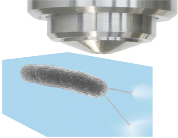
In contrast, studies reporting force quantification and mapping for bacteria are extremely scarce. This is due to the small cell size (a few µm) and low forces (~100pN) generated, which require pushing further the sensitivity limit of existing methods. The aim of this project is thus to develop a method to quantify bacterial adhesion forces. To this aim, the student will explore different photochemical and polymer physics pathway to elaborate new TFM subtrates with increased sensitivity.
Under the supervision of a postdoc working on this topic she/he will tackle the following issues:
• Synthesizing the substrates using simple click chemistry. Different routes will be explored to reach the required sensitivity.
• Benchmarking the force field measurements against readily existing methods.
• Mapping forces exerted by individual bacteria adhering on soft substrates. Our team already has experience in TFM on bacteria, and this development will be integrated in a wider project dealing with the impact of mechanical constraint on bacterial contamination of surfaces.
For whom
This 2-years master project includes microscopy, simple chemistry, image analysis and microbiology and we are thus looking for a rigorous experimentalist eager to work in an interdisciplinary environment. Some knowledge in image analysis is a plus.
It is well adapted to students of the Graduate School programm in Soft Nanosciences, following the first year major in Soft Matter and Biophysics of the master N2.
Where
The Laboratory for Interdisciplinary Physics in Grenoble brings together biophysics, soft matter physics, optics, physico-chemistry and biology in a international environment. The student will be hosted by the Laboratory of Interdisciplinary Physics (LIPhy) at UGA, in the research group Mechanics of Cells in Complex Media (MC2). He/she will work under the daily supervision of Dr Pooja Arya (postdoctoral research assistant).
Dr Delphine Débarre (microscopy, data analysis), Dr Sigolène Lecuyer (microbiology, mecanobiology) and Dr Lionel Bureau (physico-chemistry) will supervize the project.
Interested candidates should contact D. Débarre (delphine.debarre@univ-grenoble-alpes.fr) with their motivation letter, CV and transcript of university record (indicate “internship - Glycocalyx” as e-mail subject).
Published on February 12, 2021 Updated on September 7, 2022
You areYou wishSubmitShare the linkCopyCopiedClose the modal windowShare the URL of this pageI recommend:Consultable at this address:La page sera alors accessible depuis votre menu "Mes favoris".Stop videoPlay videoMutePlay audioChat: A question? Chatbot Robo FabricaMatomo traffic statisticsX (formerly Twitter)